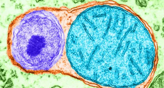
The idea of the cell as a city is a common introduction to biology, conjuring depictions of the cell’s organelles as power plants, factories, roads, libraries, warehouses and more. Like a city, these structures require a great deal of resources to build and operate, and when resources are scarce, internal components must be recycled to provide essential building blocks, particularly amino acids, to sustain vital functions.
But how do cells decide what to recycle when they are starving? One prevailing hypothesis suggests that starving cells prefer to recycle ribosomes — cellular protein-production factories rich in important amino acids and nucleotides — through autophagy, a process that degrades proteins in bulk.
However, new research by scientists at Harvard Medical School suggests otherwise. In a study published in Nature in July, they systematically surveyed the entire protein landscape of normal and nutrient-deprived cells to identify which proteins and organelles are degraded by autophagy.
The analyses revealed that, in contrast to expectations, ribosomes are not preferentially recycled through autophagy, but rather a small number of other organelles, particularly parts of the endoplasmic reticulum, are degraded.
The results shed light on how cells respond to nutrient deprivation and on autophagy and protein degradation processes, which are increasingly popular targets for drug development in cancers and other disease conditions, the authors said.
“When cells are starving, they don’t haphazardly degrade ribosomes en masse through autophagy. Instead, they appear to have mechanisms to control what they recycle,” said senior study author Wade Harper, the Bert and Natalie Vallee Professor of Molecular Pathology and chair of cell biology in the Blavatnik Institute at HMS.
“Our findings now allow us to rethink previous assumptions and better understand how cells deal with limited nutrients, a fundamental question in biology,” Harper said.
Protein turnover is a constant and universal occurrence inside every cell. To recycle unneeded or misfolded proteins, remove damaged organelles, and carry out other internal housekeeping tasks, cells utilize two primary tools, autophagy and the ubiquitin-proteasome system.
Autophagy, derived from Greek words meaning “self-eating,” allows cells to degrade proteins in bulk, as well as larger cellular structures, by engulfing them in bubble-like structures and transporting them to the cell’s waste disposal organelle, called the lysosome.
In contrast, the proteasome pathway allows cells to break down individual proteins by tagging them with a marker known as ubiquitin. Ubiquitin-modified proteins are then recognized by the proteasome and degraded.
Surprising discrepancy
Previous studies in yeast have suggested that nutrient-starved cells use autophagy to specifically recycle ribosomes, which are abundant and a reservoir of key amino acids and nucleotides. However, cells have many other mechanisms to regulate ribosome levels, and how they do so when nutrients are low has not been fully understood.
Using a combination of quantitative proteomics and genetic tools, Harper and colleagues investigated protein composition and turnover in cells that were deprived of key nutrients. To probe the role of autophagy, they also focused on cells with genetically or chemically inhibited autophagy systems.
One of the first analyses they carried out revealed that, in starving cells, total ribosomal protein levels decrease only slightly relative to other protein levels. This reduction appeared to be independent of autophagy. Cells that lacked the capacity for autophagy had no obvious defects when nutrient deprived.
“This was a very surprising finding that was at odds with existing hypotheses, and it really led us to consider that something was missing in how we think about autophagy and its role in ribosome degradation,” Harper said. “This simple result hides a huge amount of biology that we tried to uncover.”
Searching for an explanation for this discrepancy, the team, spearheaded by study co-first authors Heeseon An and Alban Ordureau, research fellows in cell biology at HMS, systematically analyzed the production of new ribosomes and the fate of existing ones in starving cells.
They did so through a variety of complementary techniques, including Ribo-Halo, which allowed them to label different ribosomal components with fluorescent tags. They could apply these tags at different time points and measure how many new ribosomes were being synthesized at the level of a single cell, as well as how many old ribosomes remained after a set amount of time.
When cells were deprived of nutrients, the primary factors that led to lower overall ribosome levels was a reduction in new ribosome synthesis and turnover through non-autophagy dependent pathways, the experiments showed. Both cell volume and the rate of cell division decreased as well, however, which allowed cells to maintain a cellular density of ribosomes.


